|
Fungal pathogen |
Apiognomonia quercina (Gleosporium asexual stage) |
|
Area(s) affected |
Leaves and twigs |
|
Signs/Symptoms |
Brown, irregular spots form on the leaves. These spots tend to follow the leaf veins and midrib causing only a portion of the leaf to turn brown. After drying, the spots will have a papery texture. Late in the growing season the fungus can be seen as dark pustules on the veins and midribs of infected leaves and dead twigs. 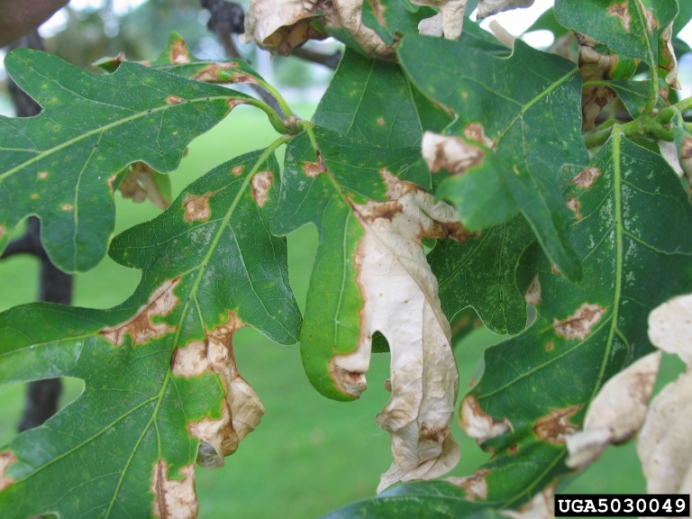 Photo credit: Joseph O’Brien, USDA Forest Service, Bugwood.org
|
|
Hosts |
Black, California black, bur, cherrybark, chestnut, English, laurel, live, California live, Oregon, pin, northern pin, post, northern red, southern red, scarlet, Shumard, water, white, swamp white and willow oaks. White oaks are the most susceptible. |
|
For more information |
http://extension.psu.edu/pests/plant-diseases/all-fact-sheets/anthracnose-on-shade-trees |
Oak Leaf Blisters
|
Fungal pathogen |
Taphrina caerulescens |
|
Area(s) affected |
Leaves |
|
Signs/Symptoms |
Occurs on species in both the red oak and white oak groups. The fungus causes leaf cells to divide abnormally and enlarge. If you flip the leaf over you will observe light-green cup-like depressions. Yellow, blister-like bulges, corresponding with the lower leaf depressions, will be seen on the upper leaf surface. As the disease progresses, blisters coalesce and will cause the leaf to curl. The fungus can sometimes be seen in the depressions, first colorless then darkens after spores are released. 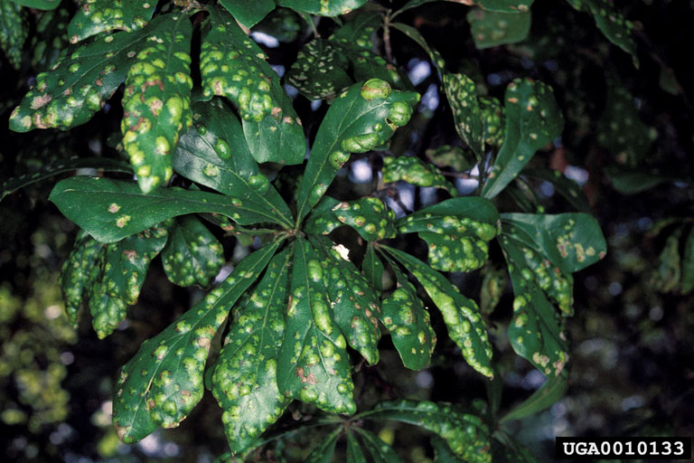 Photo credit: Andrew J. Boone, South Carolina Forestry Commission, Bugwood.org
|
|
For more information |
http://extension.psu.edu/pests/plant-diseases/all-fact-sheets/oak-leaf-blister |
Tubakia (Actinopelte) Leaf Spot
|
Fungal pathogen |
Actinopelte dryina |
|
Area(s) affected |
Leaves |
|
Signs/Symptoms |
This disease is most severe on members of the red oak group, but white oaks are also susceptible. Small, round spots form on the leaves. These spots are usually reddish-brown and tend to have a yellow halo around them. If the infection is severe, defoliation may occur. 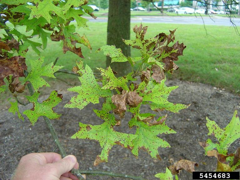 Photo credit: Jason Sharman, Vitalitree, Bugwood.org
|
|
For more information |
Spot Anthracnose
|
Fungal pathogen |
Elsinoe quercus |
|
Area(s) affected |
Leaves |
|
Signs/Symptoms |
Tiny sunken blackish-brown spots will form on the upper surface of the leaf. Symptoms are first visible in early June and increase in number until the middle of August. By that time foliage is severely damaged and the tree is a pale yellow. When infection is severe, defoliation occurs. |
|
For more information |
|
Powdery Mildew
|
Fungal pathogens |
Microsphaera alni and Phyllactinia guttata |
|
Area(s) affected |
Leaves |
|
Signs/Symptoms |
Powdery mildew occurs on all groups of oaks. Infected leaves have a faint indistinct spot on the upper leaf surface and a white to off-white powdery growth on the lower surface. The fungus will most often be found along the veins and midribs of the leaf. In severe cases, infected leaves will be slightly disfigured. 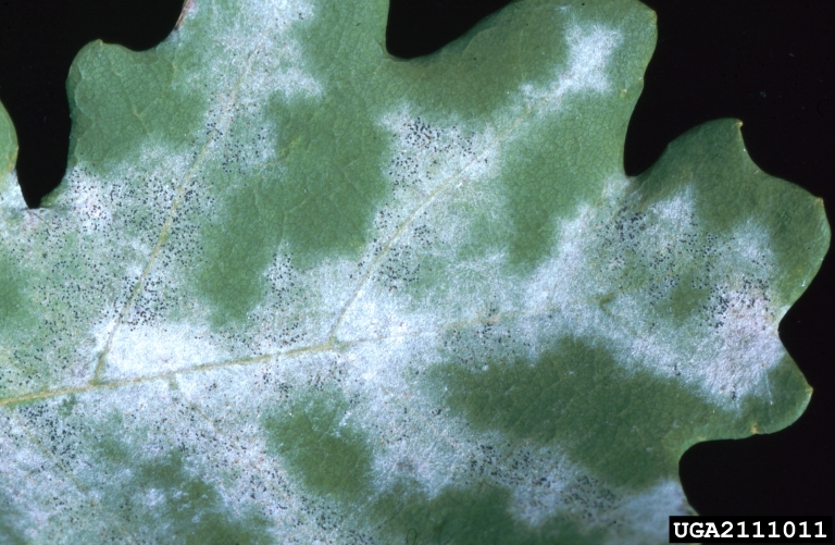 Photo credit: Petr Kapitola, State Phytosanitary Administration, Bugwood.org
|
|
For more information |
http://www.clemson.edu/extension/hgic/pests/plant_pests/flowers/hgic2049.html |
Southern Cone Rust
|
Fungal pathogen |
Cronartium strobilinum |
|
Area(s) affected |
Leaves |
|
Signs/Symptoms |
C. strobilinum infects pines in mid-winter. The telial stage is only produced on evergreen oaks, which are closely associated with live oaks. Yellow to orange pustules can be observed on the lower leaf surface of infected oak leaves. |
|
For more information |
http://www.fs.fed.us/r8/foresthealth/forestpests/diseases/socnrust.pdf |
Hypoxylon Canker
|
Fungal pathogens |
Hypoxylon atropunctatum and other Hypoxylon spp. |
|
Area(s) affected |
Entire tree |
|
Signs/Symptoms |
Hypoxylon canker occurs primarily on trees which are or have been in stressed conditions. The fungus invades the tree through injured surfaces on its limbs and trunk. The disease is first evident as a dieback of one or more branches. The foliage of the diseased limbs turns yellow and dries. This dieback continues from branch to branch until the tree dies. The fungus infects the inner bark causing the outer bark to slough off. This exposes large masses of brown, dusty spores which disseminate. Left behind is a grayish surface covered with many black fruiting bodies. The fungus causes black to brown discoloration in the sapwood which can be observed in a cross-section of the wood. 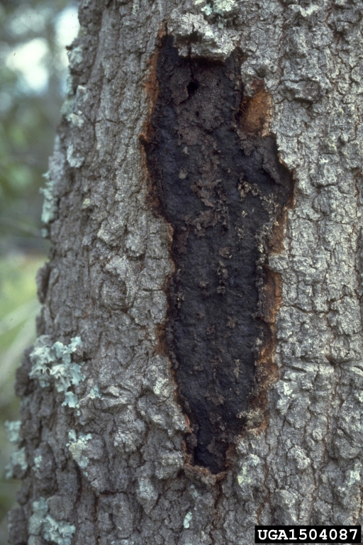 Photo credit: USDA Forest Service – Region 8 – Southern Archive, USDA Forest Service, Bugwood.org 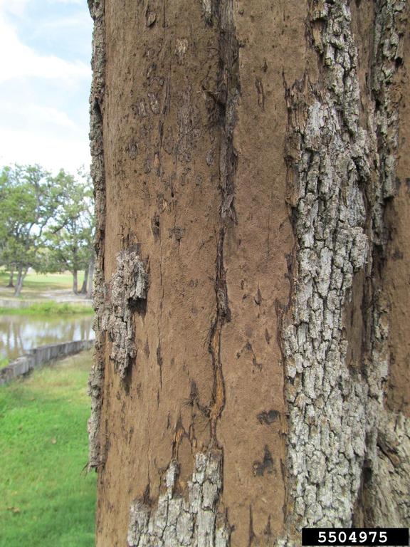 Photo credit: Molly Giesbrecht, Texas A&M AgriLife Extension Service, Bugwood.org
|
|
For more information |
http://txforestservice.tamu.edu/main/article.aspx?id=1262 http://www.fs.fed.us/r8/foresthealth/publications/r8_pr_29.pdf |
Twig Blight
|
Fungal pathogen |
Physalospora glandicola (syn. Botryosphaeria quercuum) |
|
Area(s) affected |
Twigs and branches |
|
Signs/Symptoms |
The fungus enters the tree through wounded twigs. Brown streaks can be observed in the sapwood of infected twigs and branches. Bark lesions that resemble pimple-like swellings will also develop. Infected twigs and branches die and the attached leaves will turn brown. Affected trees will look like they have been attacked by cicada. |
|
For more information |
Sinclair, Wayne A., Howard H. Lyon, and Warren T. Johnson. “Diseases of oak caused by Botryosphaeria quercuum and related fungi.” Diseases of Trees and Shrubs. Ithaca, NY: Comstock Pub. Associates, 1987. 178-79. Print. |
Endothia Canker
|
Fungal pathogen |
Endothia parasitica and Endothia gyrosa |
|
Area(s) affected |
Branches and trunks |
|
Signs/Symptoms |
Post oaks and red oaks are particularly susceptible. It has been associated with pruning cuts or limb breakage. Removal of cankers by pruning and increased tree vigor helps reduce losses. |
|
For more information |
Canker Rots
|
Fungal pathogens |
Irpex mollis, Polyporus hispidus and Poria spiculosa |
|
Area(s) affected |
Branches and trunks |
|
Signs/Symptoms |
Infections by these organisms tend to cause circular cankers which have a depressed center rather than a conk or mushroom. The center of the canker is a brown punk. They often form after a limb has been damaged or broken off.
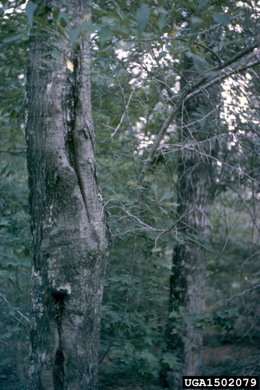 Photo credit: USDA Forest Service – Region 8 – Southern Archive, USDA Forest Service, Bugwood.org |
|
For more information |
http://www.na.fs.fed.us/spfo/pubs/fidls/canker-rots/fidl-cr.htm |
Burls
|
Fungal pathogen |
Phomopsis spp. |
|
Area(s) affected |
Branches and trunks |
|
Signs/Symptoms |
Burls can occur on both oaks and hickories. The fungus causes large tumor-like swellings on the branches and trunk of the tree. There are some swellings which occur on white oaks that aren’t caused by a fungus, but rather by the wood naturally growing over young buds and forming burl-like swellings. 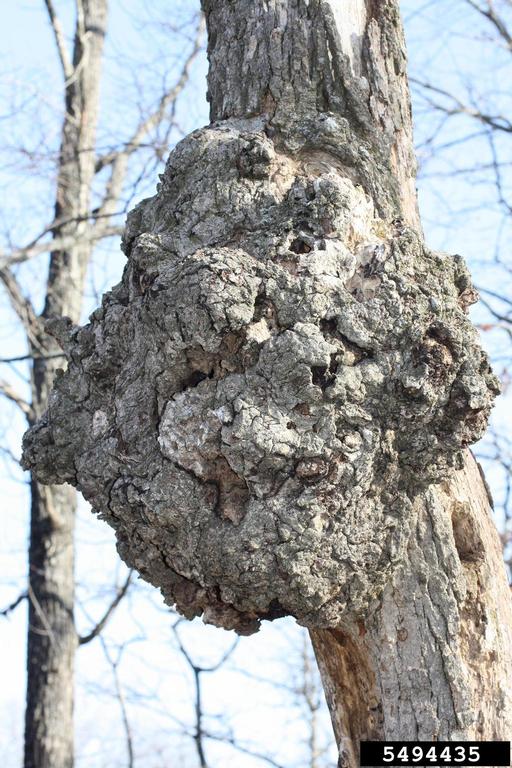 Photo credit: Robert Anderson, Bugwood.org
|
|
For more information |
http://forest.mtu.edu/research/hwbuck/hardwood_defects/burls.html |
Oak Wilt
|
Fungal pathogen |
Ceratocystis fagacearum |
|
Area(s) affected |
Entire tree |
|
Signs/Symptoms |
This is an important disease of all oaks. Members of the white oak group die very slowly after infection occurs and can carry the fungus in the vascular system without showing any symptoms. Red oaks die rapidly after infection occurs. Symptoms show up on red oaks in early May as a bronzing of the leaves. On live oak, the leaf symptoms are variable. The most common symptom is brown necrosis of the leaf veins. The remainder of the leaf may remain green or turn slightly yellow. Severe leaf drop occurs while the leaves are still green. Cuts made through the wood may show discoloration in the last annual ring. Symptom development usually begins on one limb or branch and in time spreads rapidly to the remainder of the tree. The fungus may be carried from tree to tree by various insects and through root grafts. Sap feeding beetles are important in the short range spread. Red oaks which wilt in the late summer or early fall develop spore mats under their bark during the next spring. As the mats develop, the bark sloughs off or ruptures exposing the fungus. Insects are attracted to the mats. If the insects move from mats to healthy trees which have open wounds the fungus can then enter the healthy tree and move into the water conducting tissue. 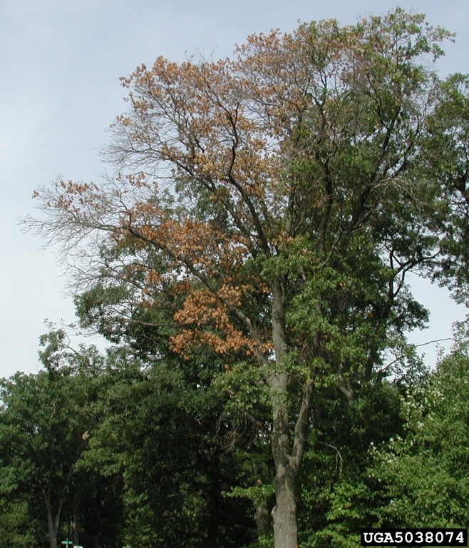 Photo credit: Joseph O’Brien, USDA Forest Service, Bugwood.org 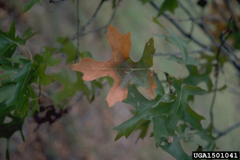 Photo credit: Paul A. Mistretta, USDA Forest Service, Bugwood.org 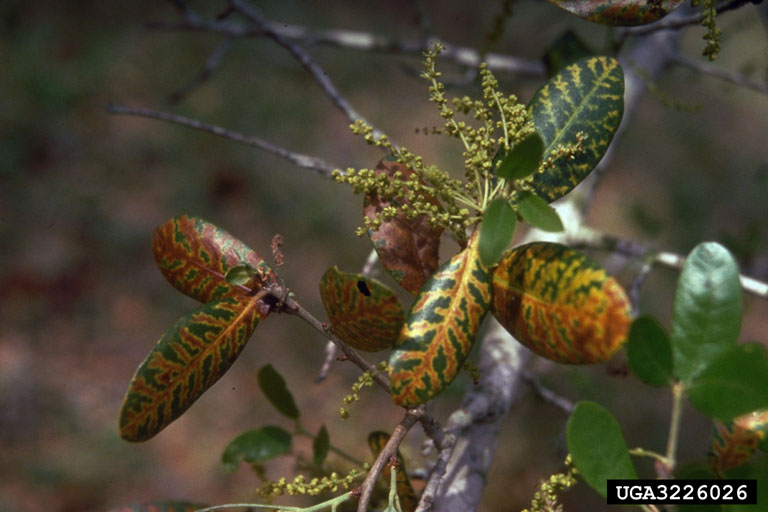 Photo credit: Ronald F. Billings, Texas Forest Service, Bugwood.org 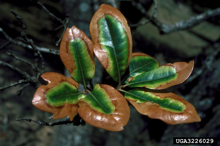 Photo credit: Ronald F. Billings, Texas Forest Service, Bugwood.org
|
|
For more information |
Oak Decline
|
Causal agents |
Combination of abiotic stress factors, fungal pathogens, and insects |
|
Area(s) affected |
Entire tree |
|
Signs/Symptoms |
Trees affected by the fungus first show signs of thinning out and twig dieback in their upper canopy. The dieback will generally increase each year. As dieback reaches the larger limbs, which comprise the main canopy, sucker growth becomes evident on the main scaffold limbs. As the disease continues to progress, only the main scaffold limbs remain alive, however, they will eventually die. This may take from five to ten years on live oaks, but less on other oaks after the first visual symptoms are observed. Live oak, post oak, water oak, Texas red oak, willow oak, sycamore, persimmon, winged elm, hackberry, American elm and western soapberry are reported to be hosts for Oak Decline. 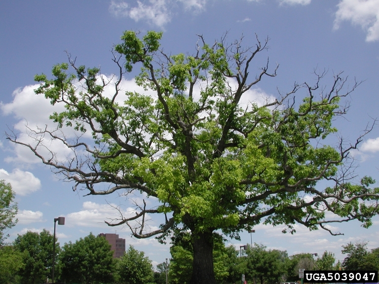 Photo credit: Joseph O’Brien, USDA Forest Service, Bugwood.org
|
|
For more information |
http://www.na.fs.fed.us/spfo/pubs/fidls/oakdecline/oakdecline.htm |
For additional support and current disease management information, contact your local AgriLife Extension Office: http://counties.agrilife.org/
Content editor: Corinne Rhodes, Undergraduate Extension Assistant, Texas Plant Disease Diagnostic Laboratory. This project was performed to satisfy BESC485 requirement under the supervision of Dr. Kevin Ong, kevo@tamu.edu, Director, Texas Plant Disease Diagnostic Laboratory, Texas A&M University, Texas AgriLife Extension Service (April 25, 2014)
