Carya illinoensis
Scab
|
Fungal pathogen |
Cladosporium caryigenum, (prev. Fusicladium effusum, Cladosporium effusum) |
|
Area(s) affected |
Leaves, nuts and green twigs |
|
Signs/Symptoms |
Small, circular, olive-green to black spots form on the lower surface of the leaf and nuts. These spots may have a velvety or cracked appearance. Sometimes these spots coalesce forming large, irregularly shaped darkened areas. On nuts, these spots appear to be sunken in. Infected twigs will exhibit elongated spots parallel to the twig axis. Infected foliage may prematurely drop. When infection is severe, the entire nut surface is black, development is arrested and the nut drops prematurely or fails to grow in the area of infection. 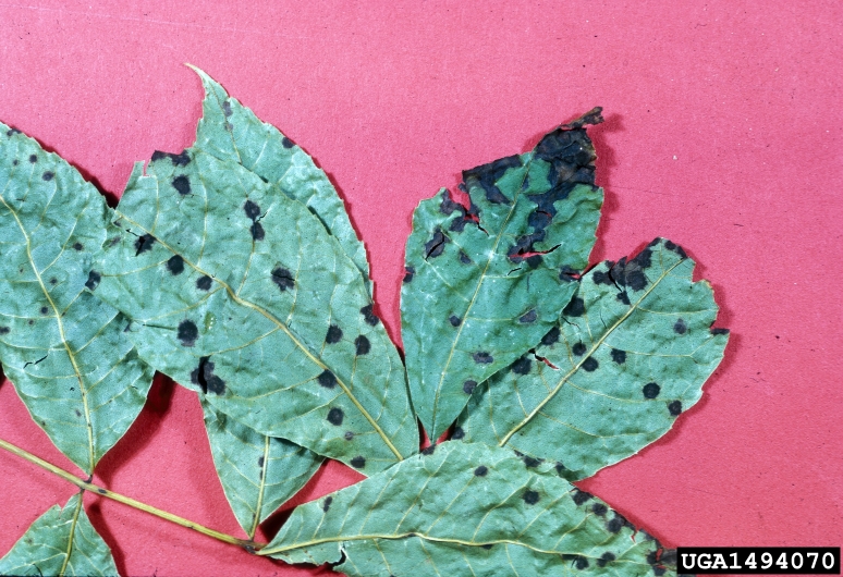 Photo credit: University of Georgia Plant Pathology Archive, University of Georgia, Bugwood.org 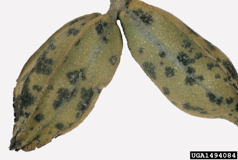 Photo credit: University of Georgia Plant Pathology Archive, University of Georgia, Bugwood.org |
|
For more information |
http://pecankernel.tamu.edu/diseases/#scab Teviotdale, Beth L., Themistocles John. Michailides, and Jay William. Pscheidt. Compendium of Nut Crop Diseases in Temperate Zones. St. Paul, MN: American Phytopathological Society, 2002. Print. |
Brown Leaf Spot
|
Fungal pathogen |
Sirosporium diffusum, (syn. Cercospora fusca) |
|
Area(s) affected |
Leaves |
|
Signs/Symptoms |
Circular, reddish brown spots form on the upper and lower leaf surface. Older spots will turn gray and concentric circles will form within the spot. If the disease is not controlled the tree will lose its leaves. |
|
For more information |
http://pecankernel.tamu.edu/diseases/#brown Teviotdale, Beth L., Themistocles John. Michailides, and Jay William. Pscheidt. Compendium of Nut Crop Diseases in Temperate Zones. St. Paul, MN: American Phytopathological Society, 2002. Print. |
Vein Spot
|
Fungal pathogen |
Gnomonia nerviseda |
|
Area(s) affected |
Leaves |
|
Signs/Symptoms |
Smooth, dark brown to black spots form on the vascular structures of the leaf which include: veins, midribs, petioles, and rachises. In the sun, these spots appear greasy or shiny. Premature defoliation will occur. This infection often resembles pecan scab lesions. |
|
For more information |
http://pecankernel.tamu.edu/diseases/#vein Teviotdale, Beth L., Themistocles John. Michailides, and Jay William. Pscheidt. Compendium of Nut Crop Diseases in Temperate Zones. St. Paul, MN: American Phytopathological Society, 2002. Print. |
Leaf Blotch
|
Fungal pathogen |
Mycosphaerella dendroides |
|
Area(s) affected |
Leaves |
|
Signs/Symptoms |
Faint yellow spots develop on the upper leaf surface; these spots eventually turn dark brown. Small, olive-green spots with a velvety appearance form on the lower leaf surface. Premature defoliation may occur. |
|
For more information |
http://pecankernel.tamu.edu/diseases/#leaf Teviotdale, Beth L., Themistocles John. Michailides, and Jay William. Pscheidt. Compendium of Nut Crop Diseases in Temperate Zones. St. Paul, MN: American Phytopathological Society, 2002. Print. |
Crown Gall
|
Bacterial pathogen |
Agrobacterium tumefaciens |
|
Area(s) affected |
Trunk and roots |
|
Signs/Symptoms |
Woody tumors or galls form on the base of the trunk and root tissue. If the gall girdles the trunk or main roots, the tree may be killed. 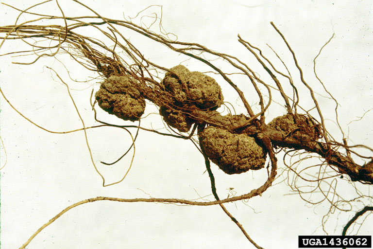 Photo credit: Clemson University – USDA Cooperative Extension Slide Series, Bugwood.org
|
|
For more information |
Teviotdale, Beth L., Themistocles John. Michailides, and Jay William. Pscheidt. Compendium of Nut Crop Diseases in Temperate Zones. St. Paul, MN: American Phytopathological Society, 2002. Print. |
Downy Spot
|
Fungal pathogen |
Mycosphaerella caryigena |
|
Area(s) affected |
Leaves |
|
Signs/Symptoms |
Circular, light yellow spots form on the lower leaf surface. These spots may be covered with a white fuzzy growth. As the disease progresses, these spots become visible on the upper leaf surface. The upper leaf surface spots are yellow, while the lower leaf surface spots turn golden brown. Premature defoliation may occur. |
|
For more information |
http://pecankernel.tamu.edu/diseases/#downy Teviotdale, Beth L., Themistocles John. Michailides, and Jay William. Pscheidt. Compendium of Nut Crop Diseases in Temperate Zones. St. Paul, MN: American Phytopathological Society, 2002. Print. |
Powdery Mildew
|
Fungal pathogen |
Microsphaera penicillata |
|
Area(s) affected |
Nuts and leaves |
|
Signs/Symptoms |
A white powdery growth develops on infected nut shucks and leaves.  Photo credit: Jerry A. Payne, USDA Agricultural Research Service, Bugwood.org
|
|
For more information |
Teviotdale, Beth L., Themistocles John. Michailides, and Jay William. Pscheidt. Compendium of Nut Crop Diseases in Temperate Zones. St. Paul, MN: American Phytopathological Society, 2002. Print. |
Pecan Bacterial Leaf Scorch (BLS)
|
Fungal pathogen(s) |
Xylella fastidiosa, Phomopsis sp., and Glomerella cingulata |
|
Area(s) affected |
Leaves |
|
Signs/Symptoms |
Brown to tan dead spots form on the margin or at the tip of the leaf. A distinct dark brown line separates the dead tissue from the living tissue. Eventually the whole leaf will turn brown and die. Premature defoliation will occur. 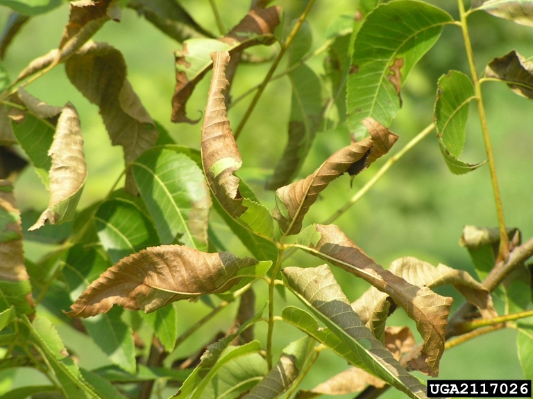 Photo credit: Rebecca A. Melanson, Louisiana State University AgCenter, Bugwood.org
|
|
For more information |
Teviotdale, Beth L., Themistocles John. Michailides, and Jay William. Pscheidt. Compendium of Nut Crop Diseases in Temperate Zones. St. Paul, MN: American Phytopathological Society, 2002. Print. |
Bunch Disease
|
Causal agent |
Phytoplasma |
|
Area(s) affected |
Shoots |
|
Signs/Symptoms |
Trees affected with bunch disease show a typical bunching symptom, caused by excessive growth of lateral buds. This results in a dense growth of thin shoots and leaves that resembles a witches’ broom. |
|
For more information |
Teviotdale, Beth L., Themistocles John. Michailides, and Jay William. Pscheidt. Compendium of Nut Crop Diseases in Temperate Zones. St. Paul, MN: American Phytopathological Society, 2002. Print. |
Parasitic and Epiphytic Plants
|
Causal agent(s) |
Fungus-algae complex, Tillandsia usneoides, Tillandsia recurvata |
|
Area(s) affected |
Trunk and branches |
|
|
Lichens: Gray to green plant that typically forms crustlike, leaflike, or branching growth on trees. 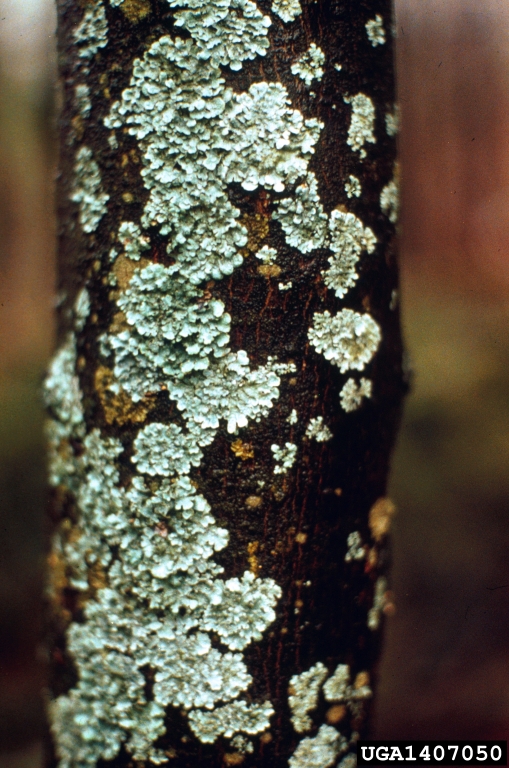 Photo credit: USDA Forest Service – Northeastern Area Archive, USDA Forest Service, Bugwood.org Spanish Moss: A long, whisker-like plant that hangs from trees. 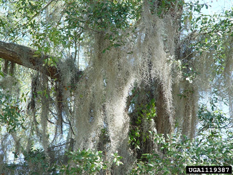 Photo credit: Wendy VanDyk Evans, Bugwood.org Ball Moss: It first occurs as small, gray green tufts that develop within a relatively short time into a dense “ball” composed of numerous individual plants. The plants form root-like holdfasts which penetrate into the rough bark of the tree. These holdfasts often completely encircle a limb. 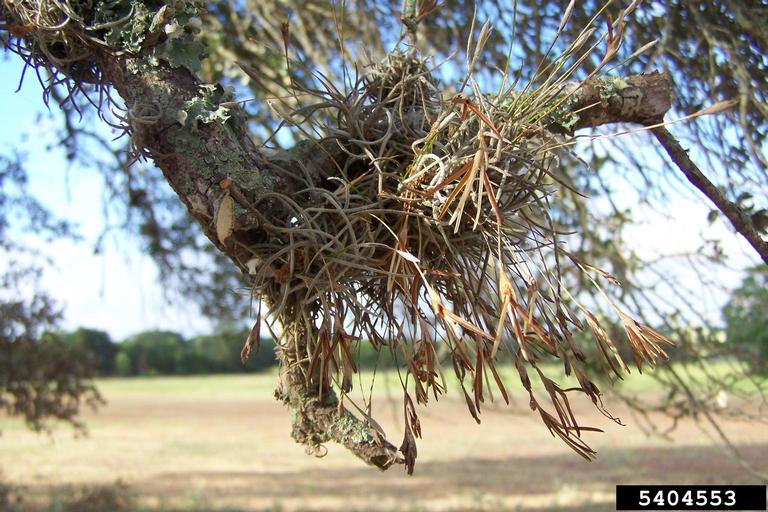 Photo credit: Karan A. Rawlins, University of Georgia, Bugwood.org |
|
For more information |
http://www.fs.fed.us/wildflowers/beauty/lichens/ |
Articularia Leaf Mold
|
Fungal pathogen |
Articularia quercina |
|
Area(s) affected |
Leaves |
|
Signs/Symptoms |
A growth of white tufts develops on the lower leaf surface. |
|
For more information |
|
Pink Mold
|
Fungal pathogen |
Trichothecium roseum |
|
Area(s) affected |
Nuts |
|
Signs/Symptoms |
Pink mold will develop on nuts that are infected with the pecan scab fungus. A white to pink, moldy growth will develop in old scab lesions. If the fungus invades the kernel, it becomes oily and produces a rancid odor. |
|
For more information |
http://pecankernel.tamu.edu/diseases/#pink
|
Cotton Root Rot
|
Fungal pathogen |
Phymatotrichopsis omnivorua, (syn. Phymatotrichum omnivorum) |
|
Area(s) affected |
Roots |
|
Signs/Symptoms |
Infected trees die suddenly. Root bark is decayed and brownish, and bronze colored wooly strands of the fungus are frequently visble on the root surface. Leaves will turn yellow or brown and will remain attached to the tree. |
|
For more information |
Teviotdale, Beth L., Themistocles John. Michailides, and Jay William. Pscheidt. Compendium of Nut Crop Diseases in Temperate Zones. St. Paul, MN: American Phytopathological Society, 2002. Print. |
Shuck Decline (Shuck Dieback)
|
Causal agent |
Unknown, possibly physiological stress or hormonal imbalance |
|
Area(s) affected |
Nuts |
|
Signs/Symptoms |
A dark, thin line forms on the inner surface of the shuck, at the junction with the shell. The inside of the shuck turns dark green and slimy and the dark, thin line progresses toward the exterior. The outer surface of the shuck has a shiny, water-soaked appearance. Beginning at the distal end, the shuck turns brown and then eventually black. The shuck may peal back at the distal end, resembling a tulip. |
|
For more information |
Teviotdale, Beth L., Themistocles John. Michailides, and Jay William. Pscheidt. Compendium of Nut Crop Diseases in Temperate Zones. St. Paul, MN: American Phytopathological Society, 2002. Print. |
Shuck and Kernel Rot
|
Fungal pathogen |
Phytophthora cactorum |
|
Area(s) affected |
Nuts |
|
Signs/Symptoms |
Fruit rot begins at the stem end of the fruit and progresses to the tip of the shuck rapidly. The shuck will turn black and become soft and moist. Kernels are inedible. |
|
For more information |
Teviotdale, Beth L., Themistocles John. Michailides, and Jay William. Pscheidt. Compendium of Nut Crop Diseases in Temperate Zones. St. Paul, MN: American Phytopathological Society, 2002. Print. |
Stem-End Blight
|
Fungal pathogen |
Unknown, suspect Botryosphaeria dothidea, (syn. Botryosphaeria ribs) |
|
Area(s) affected |
Nuts |
|
Signs/Symptoms |
Black, sunken, shiny spots form at or near the stem-end of the shuck. As the spots enlarge, the shuck will be completely black and the liquid in the kernel turns brown. |
|
For more information |
Teviotdale, Beth L., Themistocles John. Michailides, and Jay William. Pscheidt. Compendium of Nut Crop Diseases in Temperate Zones. St. Paul, MN: American Phytopathological Society, 2002. Print. |
Root Knot Nematode
|
Pathogen |
Meloidogyne spp. |
|
Area(s) affected |
Roots |
|
Signs/Symptoms |
Feed on the inside of the roots inducing knots or galls on them. These swellings are usually white and round to irregularly elongated. Foliage may wilt, appear stunted, and turn yellow or bronze. 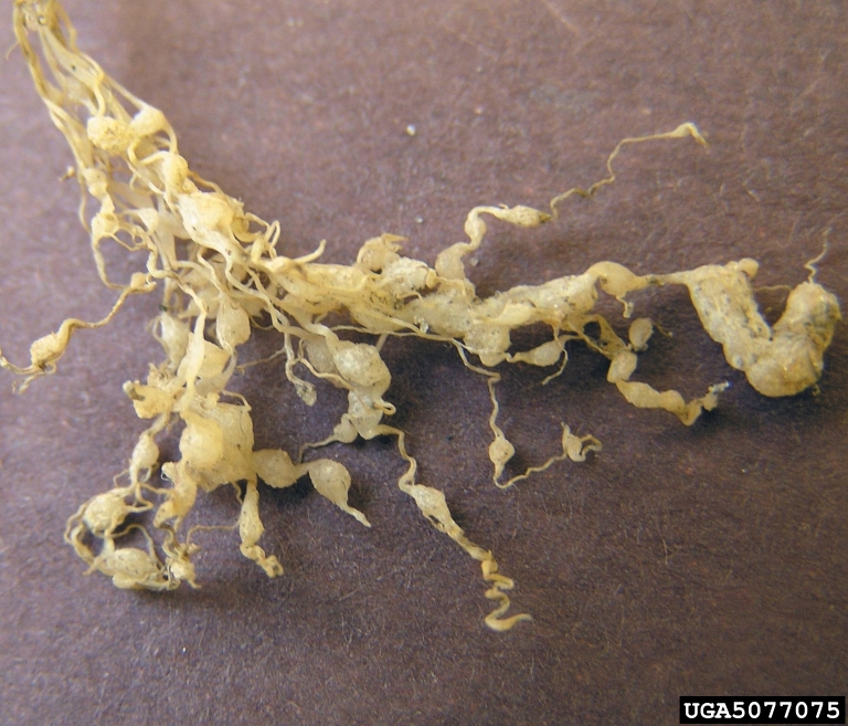 Photo credit: David B. Langston, University of Georgia, Bugwood.org
|
|
For more information |
Teviotdale, Beth L., Themistocles John. Michailides, and Jay William. Pscheidt. Compendium of Nut Crop Diseases in Temperate Zones. St. Paul, MN: American Phytopathological Society, 2002. Print. |
Kernel Discoloration
|
Fungal pathogen |
Nematospora spp. |
|
Area(s) affected |
Nuts |
|
Signs/Symptoms |
Kernels develop dark spots and may become rotted. Stinkbugs can also cause dark brown to black spots on the kernels. |
|
For more information |
Teviotdale, Beth L., Themistocles John. Michailides, and Jay William. Pscheidt. Compendium of Nut Crop Diseases in Temperate Zones. St. Paul, MN: American Phytopathological Society, 2002. Print. |
Fungal Twig Dieback
|
Fungal pathogen |
Botryosphaeria berengeriana |
|
Area(s) affected |
Branches |
|
Signs/Symptoms |
Infected branches are covered with small raised pustules with black centers. |
|
For more information |
|
For additional support and current disease management information, contact your local AgriLife Extension Office: http://counties.agrilife.org/
Content editor: Corinne Rhodes, Undergraduate Extension Assistant, Texas Plant Disease Diagnostic Laboratory. This project was performed to satisfy BESC485 requirement under the supervision of Dr. Kevin Ong, kevo@tamu.edu, Director, Texas Plant Disease Diagnostic Laboratory, Texas A&M University, Texas AgriLife Extension Service (April 25, 2014)
